Aspiration and altered airway anatomy: a presentation with a twist
Journal article, Peer reviewed
Published version
View/
Date
2018Metadata
Show full item recordCollections
- Publikasjoner fra CRIStin - SINTEF AS [5850]
- SINTEF Digital [2523]
Abstract
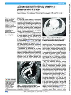
A 60-year-old woman presented to our hospital with severe hypercapnic respiratory failure in the absence of a prior smoking history. She reported a 1-day history of increased dyspnoea and a cough productive of green sputum. Her medical history was significant for severe idiopathic scoliosis, cleft palate repair, rheumatoid arthritis, hypothyroidism and recurrent lower respiratory tract infections. She was obtunded on presentation and required 24 hours of mechanical ventilation. A CT thorax was performed to further investigate her acute deterioration. Considerable distortion of thoracic anatomy secondary to kyphoscoliosis was noted. Right lower lobe atelectasis due to extrinsic compression of the bronchus intermedius by thoracic vertebrae and left lower lobe consolidation were reported (figures 1–3). Due to the vertical orientation of the left main bronchus (LMB), aspiration pneumonia was suspected (figure 2). Our patient initially improved with antimicrobial therapy, dietary modifications and proton pump inhibitors and was discharged home.